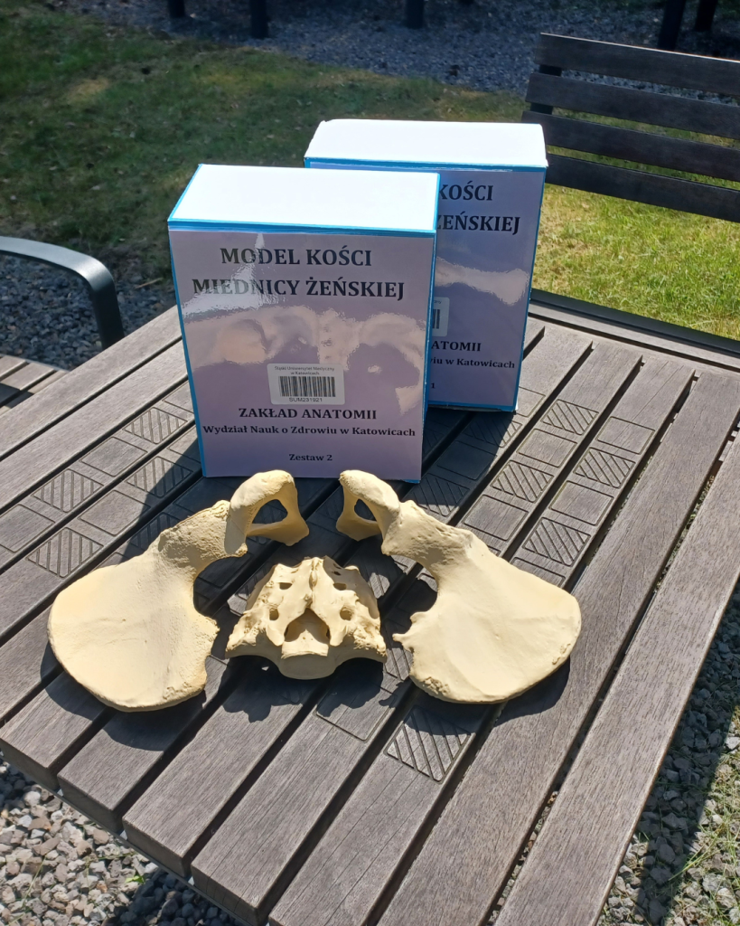
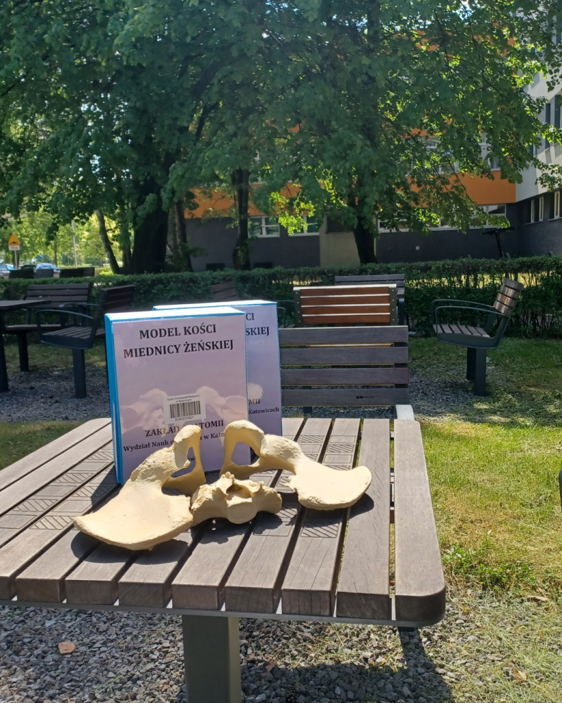
The Department of Anatomy, Faculty of Medical Sciences in Katowice, in collaboration with the Centre for Medical Education and Simulation at the Medical University of Silesia, has produced 3D prints of female pelvic bone specimens. The models were developed and printed by Łukasz Aniszewski from the Centre for Medical Education and Simulation at the Medical University of Silesia, based on real bone specimens held in the collections of the Department of Anatomy at the Faculty of Medical Sciences.
The models are designed to facilitate self-study of the pelvic bone structure, the identification of individual anatomical structures, and an understanding of the spatial orientation of these structures.
We encourage you to use the models when preparing for exams.
Models available in the reading room of the SUM Library Branch in Katowice-Ligota.
In the next sets, we’re putting together something for physiotherapists – vertebrae.